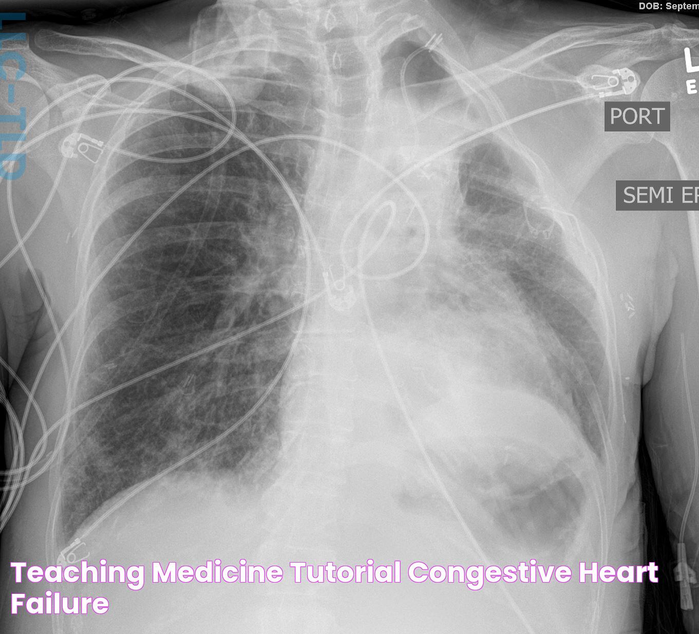
Understanding the importance of kerley c lines requires a deep dive into the anatomy of the lungs and the techniques used in radiology. These lines are more than just a part of the imaging process; they provide vital clues about the state of a patient's lungs. By examining these lines, doctors can determine if there is an abnormal accumulation of fluid or any other pulmonary complications that need to be addressed. This understanding is essential for providing optimal care to patients and improving their health outcomes.
In this comprehensive article, we will explore the intricacies of kerley c lines, from their formation to their implications in medical diagnostics. We aim to provide an in-depth understanding of their significance and how they fit into the broader context of pulmonary health. With this information, both medical professionals and patients can appreciate the role these lines play in diagnosing and treating lung conditions, leading to better healthcare experiences for all involved.
Table of Contents
- What are Kerley C Lines?
- Formation and Detection of Kerley C Lines
- Clinical Significance of Kerley C Lines
- Differentiating Kerley Lines: A, B, and C
- What Causes Kerley C Lines to Appear?
- Role of Kerley C Lines in Diagnosis
- Case Studies Involving Kerley C Lines
- How Do Radiologists Interpret Kerley C Lines?
- Advanced Imaging Techniques and Kerley C Lines
- Kerley Lines and Their Impact on Patient Care
- Common Misinterpretations and Challenges
- The Future of Kerley C Lines in Radiology
- Frequently Asked Questions About Kerley C Lines
- Conclusion
What are Kerley C Lines?
Kerley C lines are thin, linear opacities seen on chest X-rays, typically indicating interstitial pulmonary edema. Unlike Kerley A and B lines, which are more well-known, Kerley C lines are less common but still significant in diagnosing various pulmonary conditions. These lines are usually horizontal and appear at the lung bases, often indicating fluid accumulation within the interstitial spaces of the lungs.
Read also:Scott Hoying Age Biography And Insights Into His Life And Career
The presence of Kerley C lines can be a crucial diagnostic tool for radiologists and healthcare providers. They signal potential underlying conditions such as heart failure, pulmonary edema, or lymphatic congestion. Understanding these lines' appearance and implications can lead to more accurate diagnoses and better patient management strategies.
In radiological terms, Kerley C lines are indicative of the thickening of interlobular septa, which are the thin walls separating the alveolar spaces in the lungs. This thickening results from fluid or cellular infiltration, often due to increased pressure in the pulmonary veins. By recognizing these lines, healthcare professionals can better assess a patient's pulmonary health and take appropriate action.
Formation and Detection of Kerley C Lines
The formation of Kerley C lines is closely tied to the anatomy and physiology of the lungs. These lines are formed when there is an accumulation of fluid within the interstitial spaces, causing the interlobular septa to become prominent on X-rays. The increased pressure in the pulmonary veins often leads to this fluid accumulation, resulting in visible lines on imaging studies.
Detection of Kerley C lines is primarily done through chest radiographs. Radiologists look for thin, horizontal lines near the lung bases, often in conjunction with other signs of pulmonary congestion. While they are less common than other Kerley lines, their presence can provide valuable diagnostic information.
Advanced imaging techniques, such as computed tomography (CT) scans, can also aid in detecting Kerley C lines. These techniques offer a more detailed view of the lung structures, allowing for a more comprehensive assessment of the pulmonary interstitium. By utilizing these imaging modalities, healthcare professionals can gain a clearer understanding of the underlying causes of Kerley C lines and tailor their treatment approaches accordingly.
Clinical Significance of Kerley C Lines
The clinical significance of Kerley C lines lies in their ability to indicate the presence of interstitial pulmonary edema and other related conditions. These lines are often associated with heart failure, as they reflect increased pressure in the pulmonary veins due to left ventricular dysfunction. By identifying Kerley C lines, healthcare providers can better understand a patient's cardiac and pulmonary status.
Read also:Daily Nonpareil Council Bluffs Obituaries Honoring Lives And Legacies
In addition to heart failure, Kerley C lines can also indicate other conditions such as lymphatic congestion, pulmonary fibrosis, or acute respiratory distress syndrome (ARDS). Each of these conditions presents unique challenges and requires specific management strategies. Recognizing Kerley C lines can guide clinicians in their diagnostic and therapeutic approaches, ensuring that patients receive appropriate care for their underlying conditions.
Furthermore, the presence of Kerley C lines can inform treatment decisions and help monitor a patient's response to therapy. By tracking changes in these lines over time, healthcare providers can assess the effectiveness of their interventions and make necessary adjustments to optimize patient outcomes.
Differentiating Kerley Lines: A, B, and C
Kerley lines are categorized into three types: A, B, and C, each with distinct characteristics and clinical implications. Understanding the differences between these lines is crucial for accurate diagnosis and management of pulmonary conditions.
- Kerley A Lines: These are longer lines that extend from the hilum towards the lung periphery. They are often associated with pulmonary venous hypertension and indicate the redistribution of blood flow within the lungs.
- Kerley B Lines: Short, horizontal lines found at the lung bases, perpendicular to the pleura. They are indicative of interstitial edema and are the most commonly observed Kerley lines.
- Kerley C Lines: These lines are less common and represent a network of lines at the lung bases, often related to fluid accumulation in the interlobular septa.
Each type of Kerley line provides different insights into the underlying pulmonary condition, and recognizing these differences is essential for effective diagnosis and treatment. By differentiating between these lines, healthcare providers can better understand the pathophysiological processes at play and tailor their interventions to address the specific needs of their patients.
What Causes Kerley C Lines to Appear?
Kerley C lines appear due to various underlying causes that lead to the accumulation of fluid in the interstitial spaces of the lungs. The most common causes include:
- Heart Failure: Left-sided heart failure results in increased pressure in the pulmonary veins, causing fluid to leak into the interstitial spaces and leading to the formation of Kerley C lines.
- Pulmonary Edema: Fluid accumulation within the lungs due to various causes, such as cardiac or non-cardiac factors, can result in Kerley C lines appearing on chest X-rays.
- Lymphatic Congestion: Impaired lymphatic drainage can cause fluid buildup in the interlobular septa, leading to the appearance of Kerley C lines.
- Pulmonary Fibrosis: Chronic lung conditions that cause scarring and thickening of the lung tissues can also result in the formation of Kerley C lines.
Identifying the underlying cause of Kerley C lines is crucial for determining the appropriate treatment approach. By addressing the root cause, healthcare providers can effectively manage the condition and improve patient outcomes.
Role of Kerley C Lines in Diagnosis
Kerley C lines play a vital role in the diagnosis of various pulmonary conditions. Their presence on chest X-rays can provide valuable insights into the underlying health status of the lungs and the cardiovascular system. By identifying these lines, healthcare professionals can make more informed decisions regarding patient management and treatment.
In the context of heart failure, Kerley C lines can serve as an early indicator of pulmonary congestion. Their detection allows for timely intervention, potentially preventing the progression of heart failure and reducing the risk of complications. Additionally, these lines can help differentiate between cardiac and non-cardiac causes of pulmonary edema, guiding clinicians in their diagnostic process.
Furthermore, Kerley C lines can aid in the assessment of treatment efficacy. By monitoring changes in these lines over time, healthcare providers can evaluate the effectiveness of their therapeutic interventions and make necessary adjustments to optimize patient care.
Case Studies Involving Kerley C Lines
Case studies involving Kerley C lines provide valuable insights into their clinical significance and the role they play in patient management. These studies highlight real-world scenarios where the identification of Kerley C lines has led to improved diagnostic accuracy and patient outcomes.
For instance, a case study involving a patient with acute heart failure demonstrated the utility of Kerley C lines in guiding treatment decisions. The presence of these lines on the patient's chest X-ray prompted the healthcare team to initiate aggressive diuretic therapy, resulting in rapid symptom improvement and stabilization of the patient's condition.
Another case study explored the role of Kerley C lines in diagnosing lymphatic congestion in a patient with unexplained dyspnea. The identification of these lines on imaging studies led to further investigations, ultimately revealing an underlying lymphatic disorder that required targeted treatment.
These case studies underscore the importance of recognizing Kerley C lines as a valuable diagnostic tool in the context of pulmonary and cardiovascular health. By leveraging the insights gained from these lines, healthcare providers can enhance their diagnostic accuracy and deliver more effective patient care.
How Do Radiologists Interpret Kerley C Lines?
Radiologists play a crucial role in the interpretation of Kerley C lines and their implications for patient health. Their expertise in analyzing imaging studies allows them to identify these lines and assess their significance within the broader context of a patient's clinical presentation.
When interpreting Kerley C lines, radiologists consider various factors, including the patient's medical history, presenting symptoms, and other imaging findings. They evaluate the distribution and prominence of these lines, as well as any accompanying signs of pulmonary congestion or edema.
In addition to identifying Kerley C lines, radiologists collaborate with other healthcare professionals to determine the underlying cause of these lines and develop an appropriate treatment plan. This multidisciplinary approach ensures that patients receive comprehensive care that addresses their specific needs.
Advanced Imaging Techniques and Kerley C Lines
Advanced imaging techniques have revolutionized the detection and assessment of Kerley C lines, providing healthcare professionals with more detailed and accurate information about a patient's pulmonary status. These techniques include computed tomography (CT) scans and magnetic resonance imaging (MRI), which offer enhanced visualization of the lung structures and interstitial spaces.
CT scans, in particular, are highly effective in identifying Kerley C lines and evaluating their significance within the context of a patient's clinical presentation. These scans provide cross-sectional images of the chest, allowing for a more comprehensive assessment of the pulmonary interstitium and any associated abnormalities.
By utilizing advanced imaging techniques, healthcare providers can gain a deeper understanding of the underlying causes of Kerley C lines and develop more targeted treatment strategies. This approach enhances diagnostic accuracy and improves patient outcomes, ultimately leading to better healthcare experiences for patients.
Kerley Lines and Their Impact on Patient Care
The presence of Kerley lines, including Kerley C lines, has a significant impact on patient care and management. These lines provide valuable insights into a patient's pulmonary and cardiovascular health, guiding healthcare providers in their diagnostic and therapeutic approaches.
By identifying Kerley lines on imaging studies, healthcare professionals can make more informed decisions regarding patient management. These lines serve as early indicators of pulmonary congestion and other related conditions, allowing for timely intervention and prevention of complications.
Moreover, Kerley lines play a crucial role in monitoring treatment efficacy. By tracking changes in these lines over time, healthcare providers can assess the effectiveness of their interventions and make necessary adjustments to optimize patient outcomes.
Overall, the recognition and understanding of Kerley lines, including Kerley C lines, are essential for delivering high-quality patient care. By leveraging the insights gained from these lines, healthcare professionals can enhance their diagnostic accuracy and improve patient outcomes.
Common Misinterpretations and Challenges
While Kerley C lines provide valuable diagnostic information, their interpretation can present challenges and potential misinterpretations. Radiologists and healthcare providers must be aware of these challenges to ensure accurate diagnosis and effective patient management.
One common challenge is differentiating Kerley C lines from other similar-appearing lines or shadows on chest X-rays. Misinterpretation of these lines can lead to incorrect diagnoses and inappropriate treatment strategies. To address this challenge, radiologists must consider the patient's clinical presentation and other imaging findings when interpreting Kerley C lines.
Another challenge is the potential for false-positive results, where Kerley C lines are identified in the absence of significant pulmonary or cardiovascular pathology. In such cases, further investigations may be necessary to confirm the underlying cause and guide appropriate management.
By recognizing these challenges and potential misinterpretations, healthcare professionals can enhance their diagnostic accuracy and ensure that patients receive appropriate care for their specific conditions.
The Future of Kerley C Lines in Radiology
The future of Kerley C lines in radiology is promising, with advancements in imaging technology and diagnostic techniques continuing to enhance their detection and interpretation. These developments are expected to improve the accuracy and efficiency of diagnosing pulmonary and cardiovascular conditions, ultimately leading to better patient outcomes.
Emerging technologies, such as artificial intelligence (AI) and machine learning, are poised to play a significant role in the future of Kerley C lines. These technologies have the potential to automate the detection and analysis of Kerley lines, reducing the risk of human error and improving diagnostic consistency.
Additionally, ongoing research into the pathophysiology and clinical implications of Kerley C lines is expected to expand our understanding of these lines and their role in patient care. This research will inform the development of new diagnostic and therapeutic strategies, ensuring that healthcare providers can deliver the highest quality care to their patients.
Overall, the future of Kerley C lines in radiology holds great potential for advancing the field and improving patient outcomes. By embracing these advancements, healthcare professionals can enhance their diagnostic accuracy and deliver more effective care to their patients.
Frequently Asked Questions About Kerley C Lines
- What are Kerley C lines? Kerley C lines are thin, linear opacities seen on chest X-rays, typically indicating interstitial pulmonary edema. They are less common than Kerley A and B lines but still significant in diagnosing various pulmonary conditions.
- How are Kerley C lines detected? Kerley C lines are primarily detected through chest radiographs. Advanced imaging techniques, such as computed tomography (CT) scans, can also aid in their detection, offering a more detailed view of the lung structures.
- What causes Kerley C lines to appear? Kerley C lines can appear due to various underlying causes, including heart failure, pulmonary edema, lymphatic congestion, and pulmonary fibrosis. Each of these conditions leads to fluid accumulation in the interstitial spaces of the lungs.
- What is the clinical significance of Kerley C lines? The clinical significance of Kerley C lines lies in their ability to indicate the presence of interstitial pulmonary edema and other related conditions. They are often associated with heart failure and provide valuable diagnostic information for healthcare providers.
- How do radiologists interpret Kerley C lines? Radiologists interpret Kerley C lines by analyzing imaging studies and considering the patient's medical history, presenting symptoms, and other imaging findings. They collaborate with other healthcare professionals to determine the underlying cause and develop an appropriate treatment plan.
- What is the future of Kerley C lines in radiology? The future of Kerley C lines in radiology is promising, with advancements in imaging technology and diagnostic techniques continuing to enhance their detection and interpretation. Emerging technologies, such as artificial intelligence, are expected to play a significant role in improving diagnostic accuracy and patient outcomes.
Conclusion
Kerley C lines, although less commonly discussed than their counterparts, hold significant importance in the field of radiology and pulmonary diagnostics. Their presence on chest X-rays provides critical insights into a patient's pulmonary health, aiding in the diagnosis and management of various conditions. Through a comprehensive understanding of these lines, healthcare professionals can enhance their diagnostic accuracy, leading to improved patient outcomes.
The future of Kerley C lines in radiology is bright, with ongoing advancements in imaging technology and diagnostic techniques poised to further enhance their utility. By embracing these developments, healthcare providers can deliver more effective and efficient care, ultimately improving the health and well-being of their patients.
As we continue to explore the intricacies of Kerley C lines and their implications in patient care, it is essential to remain informed and adaptable. By staying abreast of the latest research and technological advancements, healthcare professionals can ensure they are providing the highest quality care to their patients, ultimately leading to better health outcomes and improved quality of life.

