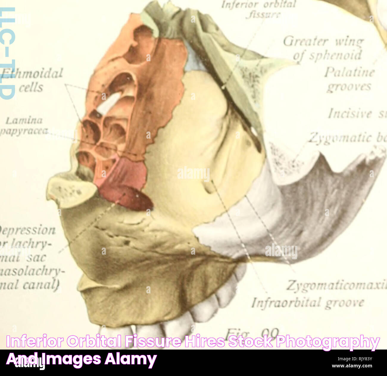
The importance of the inferior orbital fissure extends beyond its anatomical placement. It serves as a conduit for several essential structures, including nerves and blood vessels that supply the orbit and surrounding areas. This fissure is also integral in maintaining the communication between the orbit and the infratemporal and pterygopalatine fossae. The anatomical relationships and structures associated with the inferior orbital fissure make it a subject of interest for both medical professionals and anatomy enthusiasts. In medical practice, knowledge about the inferior orbital fissure is vital, especially for specialists in ophthalmology and neurology. Its significance is underscored in various clinical scenarios, such as orbital fractures, nerve entrapments, and surgical approaches to the orbit. A comprehensive understanding of this feature can aid in diagnosing and managing conditions that affect the eye and the surrounding regions. This article delves into the complexities of the inferior orbital fissure, exploring its anatomy, function, and clinical relevance.
Table of Contents
1. What is the Anatomy of the Inferior Orbital Fissure? 2. Which Structures Pass Through the Inferior Orbital Fissure? 3. Where is the Inferior Orbital Fissure Located? 4. What is the Function of the Inferior Orbital Fissure? 5. Why is the Inferior Orbital Fissure Clinically Significant? 6. How Does the Inferior Orbital Fissure Develop? 7. What Are the Variants of the Inferior Orbital Fissure? 8. Can Disorders Affect the Inferior Orbital Fissure? 9. How is the Inferior Orbital Fissure Diagnosed? 10. What Treatments Involve the Inferior Orbital Fissure? 11. What Surgical Approaches Involve the Inferior Orbital Fissure? 12. What Does Research Say About the Inferior Orbital Fissure? 13. Frequently Asked Questions 14. Conclusion
What is the Anatomy of the Inferior Orbital Fissure?
The inferior orbital fissure is an intricate anatomical structure located at the juncture of the greater wing of the sphenoid bone and the maxilla. This fissure forms an integral part of the eye socket, or orbit, acting as a critical passageway for nerves and blood vessels. Its elongated shape and strategic location underscore its importance in linking various cranial structures.
Read also:Affordable Streaming With Fire Tv Stick Open Box Canada
The fissure is bordered superiorly by the greater wing of the sphenoid bone and inferiorly by the orbital process of the maxilla. Laterally, it is flanked by the zygomatic bone, while medially, it approaches the body of the sphenoid. This positioning within the orbit facilitates its role as a conduit for several neurovascular structures.
In terms of dimension, the inferior orbital fissure's size can vary among individuals, but it generally measures approximately 15 to 25 millimeters in length. This variance is attributed to genetic and developmental factors, indicating the complexity and individuality of human anatomy. Despite these variations, the fissure consistently maintains its role in facilitating communication between the orbit and adjacent fossae.
Which Structures Pass Through the Inferior Orbital Fissure?
The inferior orbital fissure serves as a vital passageway for numerous structures. Primarily, it allows the passage of the infraorbital nerve, a branch of the maxillary nerve (CN V2), which provides sensory innervation to the upper cheek, lower eyelid, and upper lip. Additionally, the zygomatic nerve, another branch of the maxillary nerve, traverses this fissure, contributing to the sensory innervation of the face.
Moreover, the inferior orbital fissure accommodates the infraorbital artery and vein. These vascular structures are responsible for supplying blood to and draining blood from the midfacial region, respectively. The artery originates from the maxillary artery, while the vein eventually drains into the pterygoid venous plexus.
Other structures that pass through the inferior orbital fissure include the inferior ophthalmic vein, which plays a crucial role in draining venous blood from the orbit. This vein's passage through the fissure facilitates its communication with the pterygoid plexus and the cavernous sinus, highlighting the fissure's importance in orbital venous drainage.
Where is the Inferior Orbital Fissure Located?
The inferior orbital fissure is strategically positioned at the base of the orbit, serving as a gateway between the orbit and surrounding anatomical regions. This location is pivotal in its function as a conduit for nerves and vessels that connect the orbit to other cranial structures.
Read also:All You Need To Know About Pensacola Pediatrics For Your Childs Health
Geographically, the inferior orbital fissure is located in the anterior cranial fossa, specifically at the junction of the greater wing of the sphenoid bone and the maxilla. It lies beneath the superior orbital fissure, another critical anatomical feature, and is oriented horizontally.
The fissure's proximity to the maxillary sinus and the pterygopalatine fossa further emphasizes its anatomical significance. This location facilitates the transmission of neurovascular structures between the orbit and these adjacent regions, underscoring the fissure's role in cranial and facial connectivity.
What is the Function of the Inferior Orbital Fissure?
The primary function of the inferior orbital fissure is to serve as a passageway for essential neurovascular structures. These structures include the infraorbital nerve, zygomatic nerve, and the infraorbital artery and vein, all of which are vital for sensory innervation and blood supply to the midface.
Beyond its role in transmitting neurovascular structures, the inferior orbital fissure plays a critical part in maintaining communication between the orbit and surrounding fossae. This communication is crucial for the balance and coordination of sensory and motor signals within the cranial cavity, highlighting the fissure's role in the overall function of the head and face.
The fissure's orientation and location also contribute to its function. By linking the orbit to adjacent anatomical regions, the inferior orbital fissure facilitates the integration of sensory and vascular networks, ensuring efficient transmission of signals and resources throughout the cranium.
Why is the Inferior Orbital Fissure Clinically Significant?
The clinical significance of the inferior orbital fissure is underscored in various medical scenarios. Its role as a conduit for neurovascular structures makes it a focal point in the diagnosis and management of orbital and facial conditions.
In cases of orbital fractures, the inferior orbital fissure can be affected, leading to nerve entrapments or vascular disruptions. Understanding its anatomy and function aids clinicians in diagnosing such injuries and planning appropriate surgical interventions to restore normal function.
Moreover, the fissure's involvement in transmitting the infraorbital nerve and artery makes it a critical area in procedures related to anesthesia and pain management. Knowledge of its location and the structures it houses is essential for administering effective regional anesthesia in the midface region.
How Does the Inferior Orbital Fissure Develop?
The development of the inferior orbital fissure is a complex process that occurs during craniofacial development in the embryonic stage. It involves the interaction and growth of various cranial bones, including the sphenoid, maxilla, and zygomatic bones.
Initially, the orbit and its structures form from the mesenchymal tissue, which differentiates into the skeletal components of the skull. As the bones develop and fuse, the inferior orbital fissure emerges as a separation between the greater wing of the sphenoid bone and the maxilla.
This developmental process is influenced by genetic factors and signaling pathways that regulate bone growth and differentiation. Any disruptions in these pathways can result in variations or anomalies in the formation of the inferior orbital fissure, which may have clinical implications.
What Are the Variants of the Inferior Orbital Fissure?
Anatomical variations of the inferior orbital fissure are not uncommon and can be attributed to genetic and developmental factors. These variants may involve differences in the size, shape, or orientation of the fissure, affecting its function and the structures that pass through it.
Some individuals may exhibit a wider or narrower fissure, which can impact the transmission of neurovascular structures. Additionally, the presence of accessory fissures or foramina in the vicinity of the inferior orbital fissure can alter the distribution of nerves and vessels, potentially affecting sensory and vascular supply.
While these variations are generally benign, they can have clinical significance in certain situations, such as surgical planning or the interpretation of imaging studies. Understanding these anatomical differences is crucial for healthcare professionals involved in the diagnosis and treatment of orbital and facial conditions.
Can Disorders Affect the Inferior Orbital Fissure?
Yes, various disorders can impact the inferior orbital fissure, leading to clinical symptoms and requiring medical intervention. These disorders may arise from trauma, congenital anomalies, or pathological processes affecting the orbit and surrounding structures.
Orbital fractures, for instance, can involve the inferior orbital fissure, resulting in nerve entrapment or vascular injury. This can lead to symptoms such as facial numbness, pain, or visual disturbances, necessitating surgical repair to restore normal function.
In some cases, tumors or lesions in the orbit or adjacent fossae may impinge upon the inferior orbital fissure, compressing the structures within it. This can result in sensory deficits or vascular compromise, requiring prompt diagnosis and treatment to prevent further complications.
How is the Inferior Orbital Fissure Diagnosed?
The diagnosis of conditions affecting the inferior orbital fissure often involves a combination of clinical assessment and imaging studies. A thorough understanding of its anatomy and function is essential for accurate diagnosis and effective management.
Imaging techniques such as computed tomography (CT) and magnetic resonance imaging (MRI) are commonly used to visualize the inferior orbital fissure and assess any abnormalities. These modalities provide detailed images of the bony structures and soft tissues, aiding in the identification of fractures, lesions, or anatomical variations.
In addition to imaging, clinical evaluation plays a crucial role in diagnosing conditions related to the inferior orbital fissure. A comprehensive assessment of symptoms, such as facial numbness or pain, along with a detailed medical history, can provide valuable insights into the underlying cause and guide further diagnostic investigations.
What Treatments Involve the Inferior Orbital Fissure?
Treatment approaches for conditions involving the inferior orbital fissure vary depending on the underlying cause and the structures affected. Surgical intervention is often necessary in cases of trauma or tumors, aiming to restore normal anatomy and function.
In instances of orbital fractures involving the inferior orbital fissure, surgical repair may be required to realign the bones and release any entrapped nerves or vessels. This can help alleviate symptoms such as pain, numbness, or visual disturbances, improving the patient's quality of life.
For tumors or lesions impacting the inferior orbital fissure, surgical excision or debulking may be performed to relieve pressure on the affected structures. This can help restore normal sensory and vascular function, preventing further complications.
What Surgical Approaches Involve the Inferior Orbital Fissure?
Various surgical approaches involve the inferior orbital fissure, particularly in the context of orbital and craniofacial surgeries. These approaches are designed to access the orbit and surrounding structures, allowing for the treatment of fractures, tumors, or other pathological conditions.
One common approach is the transconjunctival approach, which involves an incision in the lower eyelid to access the orbit and inferior orbital fissure. This technique provides excellent exposure while minimizing visible scars, making it a preferred option for cosmetic and functional procedures.
Another approach is the transantral approach, which involves accessing the orbit through the maxillary sinus. This technique is often used in the treatment of orbital floor fractures or lesions involving the inferior orbital fissure, providing direct access to the affected area.
What Does Research Say About the Inferior Orbital Fissure?
Research on the inferior orbital fissure continues to expand our understanding of its anatomy, function, and clinical relevance. Ongoing studies aim to elucidate the developmental pathways and genetic factors that influence its formation and variations.
Recent advancements in imaging techniques have enhanced our ability to visualize the inferior orbital fissure and assess its role in various clinical conditions. These developments have improved diagnostic accuracy and informed surgical approaches, contributing to better patient outcomes.
Additionally, research into the neurovascular structures associated with the inferior orbital fissure has provided insights into their function and potential implications for disorders such as trigeminal neuralgia or vascular anomalies. This knowledge is essential for developing targeted treatments and improving our understanding of craniofacial anatomy.
Frequently Asked Questions
What is the inferior orbital fissure?
The inferior orbital fissure is an anatomical opening located at the base of the orbit, facilitating the passage of nerves and blood vessels between the orbit and adjacent cranial structures.
What structures pass through the inferior orbital fissure?
Structures passing through the inferior orbital fissure include the infraorbital nerve, zygomatic nerve, infraorbital artery, infraorbital vein, and inferior ophthalmic vein.
How is the inferior orbital fissure diagnosed?
Diagnosis often involves imaging techniques such as CT and MRI, along with clinical assessment to evaluate symptoms and identify any abnormalities affecting the fissure.
Can disorders affect the inferior orbital fissure?
Yes, disorders such as orbital fractures, tumors, or congenital anomalies can impact the inferior orbital fissure, leading to symptoms that may require medical intervention.
Why is the inferior orbital fissure clinically significant?
Its role as a conduit for neurovascular structures makes it significant in the diagnosis and treatment of orbital and facial conditions, particularly in trauma and surgical procedures.
What surgical approaches involve the inferior orbital fissure?
Surgical approaches such as the transconjunctival and transantral techniques are used to access the orbit and treat conditions involving the inferior orbital fissure.
Conclusion
In conclusion, the inferior orbital fissure is a vital component of human anatomy, playing a crucial role in the function and connectivity of the orbit and surrounding structures. Its significance in clinical practice is underscored by its involvement in various conditions and surgical procedures. Understanding the anatomy, function, and clinical relevance of the inferior orbital fissure is essential for healthcare professionals involved in the diagnosis and treatment of orbital and craniofacial conditions. As research continues to advance, our knowledge of this intricate anatomical feature will undoubtedly expand, leading to improved patient care and outcomes.
For more in-depth information on the inferior orbital fissure and its clinical implications, you can explore resources such as NCBI and other reputable medical literature.

